
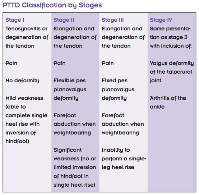
In summary, custom foot orthotics with adequate rearfoot control will be beneficial in patients with flexible sub-talar joints (stages 1 – 2). In patients with rigid planovalgus foot deformity (or absence of a spring ligament due to rupture), the benefits of custom foot orthotics is considered limited.
Key considerations when treating PTTD stages 1, 2a and 2b:
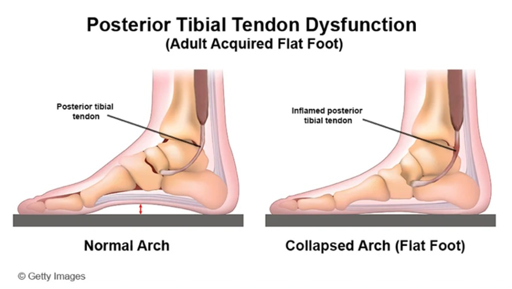
As noted above, patients with stages 1 and 2 PTTD will present with a flexible, planus foot type. As such, the key to effective management is an adequate rearfoot varus posting and full MLA contour – ensuring the suspected supinutus is removed during your 3D scanning.
Secondly, in patients with a large midfoot bulge, or prominent navicular, increasing the heel or midfoot width with a flange can be a beneficial way to ensure the patient has full contour through the rearfoot and midfoot. If this area is painful, applying a dell and poron filling the specific area (marked on your 3D scan for accuracy) and further offload the tissue.